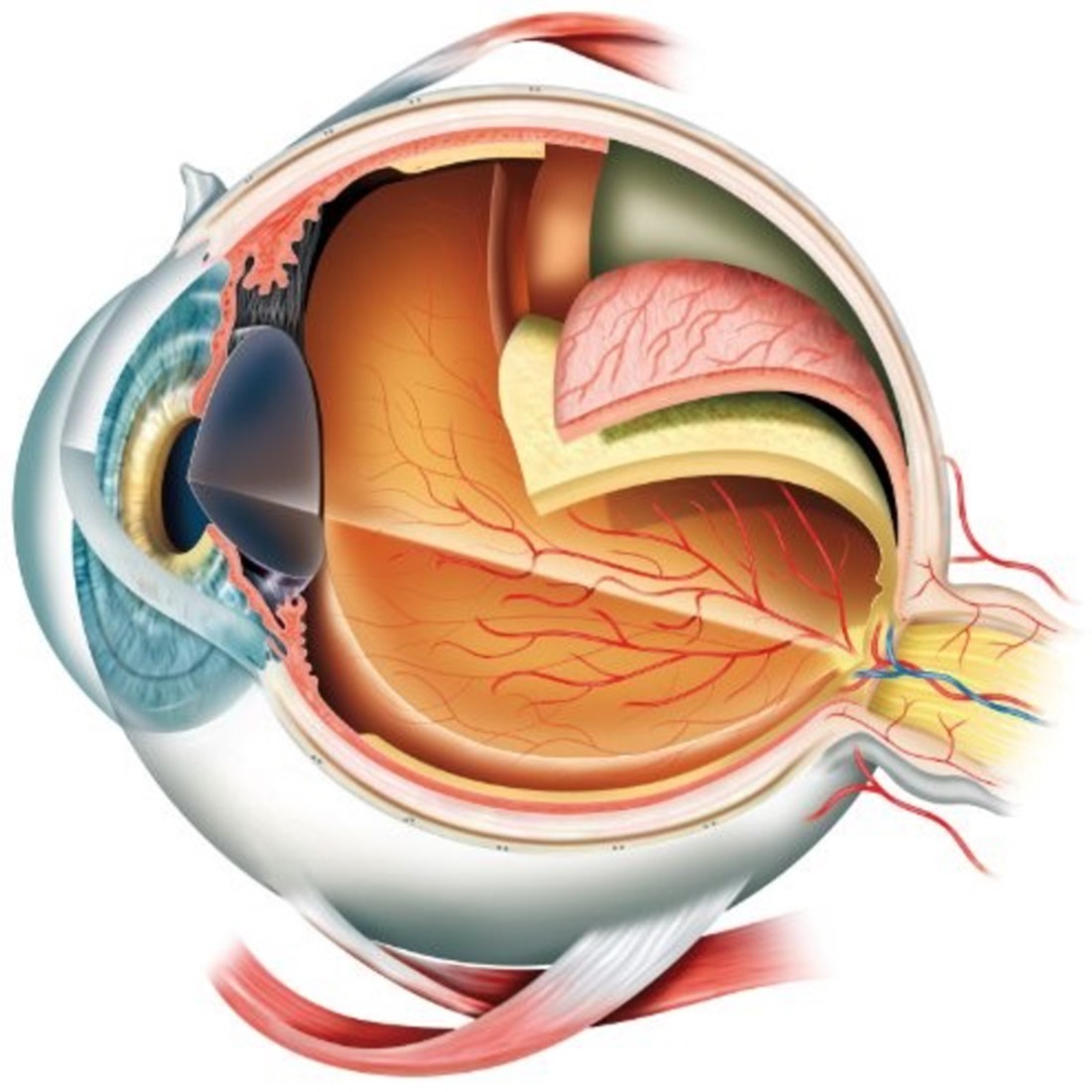
The Anatomy of the Eye
The human eye is a complex and intricate organ, essential for vision. Understanding its anatomy is crucial for diagnosing and treating various ocular conditions. This summary provides a summary of the key structures of the eye and their functions.

External Structures
Eyelids and Eyelashes
The eyelids protect the eye from foreign particles, excessive light, and injury. They also help spread tears over the surface of the eye to keep it moist. Eyelashes act as a barrier to dust and debris.
Conjunctiva
The conjunctiva is a thin, transparent membrane that covers the white part of the eye (sclera) and the inner surfaces of the eyelids. It helps lubricate the eye by producing mucus and tears.
Lacrimal Apparatus
The lacrimal apparatus consists of the lacrimal glands, which produce tears, and the lacrimal ducts, which drain tears into the nasal cavity. Tears are essential for keeping the eye moist, providing nutrients, and protecting against infection.
Internal Structures
Cornea
The cornea is the eye's outermost layer, a clear, dome-shaped surface that covers the front of the eye. It plays a crucial role in focusing light onto the retina. The cornea is composed of five layers: the epithelium, Bowman's layer, stroma, Descemet's membrane, and endothelium.
Sclera
The sclera is the white, opaque part of the eye that provides structural support and protection. It is continuous with the cornea at the front and extends to the optic nerve at the back.
Anterior Chamber
The anterior chamber is the fluid-filled space between the cornea and the iris. It contains aqueous humor, a clear fluid that nourishes the cornea and lens and maintains intraocular pressure.
Iris and Pupil
The iris is the colored part of the eye that controls the size of the pupil, the opening in the center of the iris. The iris adjusts the pupil size to regulate the amount of light entering the eye.
Lens
The lens is a transparent, biconvex structure located behind the iris. It focuses light onto the retina by changing its shape, a process known as accommodation. The lens is held in place by the zonules, fine fibers that connect it to the ciliary body.
Ciliary Body
The ciliary body is a ring of tissue that encircles the lens. It contains the ciliary muscle, which controls the shape of the lens, and the ciliary processes, which produce aqueous humor.
Choroid
The choroid is a layer of blood vessels and connective tissue between the sclera and the retina. It provides oxygen and nutrients to the outer layers of the retina.
Retina
The retina is the light-sensitive layer at the back of the eye. It contains photoreceptor cells (rods and cones) that convert light into electrical signals. Rods are responsible for vision in low light, while cones are responsible for color vision and detail. The retina also contains several layers of neurons that process visual information before sending it to the brain via the optic nerve.
Macula and Fovea
The macula is a small, central area of the retina responsible for detailed central vision. The fovea, located in the center of the macula, contains a high concentration of cones and is the point of sharpest vision.
Optic Nerve
The optic nerve is a bundle of over a million nerve fibers that transmit visual information from the retina to the brain. It exits the eye at the optic disc, a small, circular area where there are no photoreceptors, creating a natural blind spot.
Vitreous Body
The vitreous body is a clear, gel-like substance that fills the space between the lens and the retina. It helps maintain the eye's shape and allows light to pass through to the retina.
Blood Supply
The eye receives blood from the ophthalmic artery, a branch of the internal carotid artery. The central retinal artery supplies the inner retina, while the ciliary arteries supply the outer retina, choroid, and other structures.
Nerve Supply
The eye's sensory innervation is provided by the ophthalmic branch of the trigeminal nerve (cranial nerve V). The optic nerve (cranial nerve II) transmits visual information to the brain. The oculomotor (cranial nerve III), trochlear (cranial nerve IV), and abducens (cranial nerve VI) nerves control the eye muscles.
Eye Muscles
The eye is moved by six extraocular muscles: the superior, inferior, medial, and lateral rectus muscles, and the superior and inferior oblique muscles. These muscles are controlled by the oculomotor, trochlear, and abducens nerves.
Superior Rectus
The superior rectus elevates the eye and turns it medially.
Inferior Rectus
The inferior rectus depresses the eye and turns it medially.
Medial Rectus
The medial rectus moves the eye medially.
Lateral Rectus
The lateral rectus moves the eye laterally.
Superior Oblique
The superior oblique depresses the eye and turns it laterally.
Inferior Oblique
The inferior oblique elevates the eye and turns it laterally.
Conclusion
The eye is a remarkable organ with a complex structure that allows us to perceive the world in vivid detail. Understanding its anatomy is essential for diagnosing and treating ocular conditions. By appreciating the intricate design and function of each part of the eye, ophthalmologists can provide better care and improve patients' quality of life.

